Knee problems are a huge frustration for many, from competitive athletes to those who simply enjoy gardening, and can place unwanted restrictions on your daily life.
There are many causes, but accurate management of the problem relies upon accurate diagnosis.
Osteoarthritis (OA)
A degenerative condition commonly known as 'wear and tear' of a joint. OA typically affects those over 45 and produces a pattern of fluctuating stiffness, swelling and pain. 'Wear and tear' can develop gradually over the years as a result of overuse of a joint, where the protective hyaline cartilage within the joint gradually becomes worn and the joint surfaces are left to effectively 'rub' on one another. Trauma to the joint at an earlier stage of an individual's life is thought to predispose them to earlier degeneration within that joint. Injuries such as ligament damage (for example, ACL injury) may show earlier joint changes on xray / MRI scan than the uninjured side due to the altered joint mechanics and stresses that are produced within the knee joint following such an injury.
It's important to mention that not all individuals with degenerative changes within their joints experience pain or any other symptoms, and therefore osteoarthritis affects people differently.
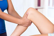 Although there is no cure for osteoarthritis and it is a progressive disease of the joint, there are certain management strategies that can improve the symptoms, and indeed prolong the integrity of the joint. A well balanced diet, cod liver oil supplements and adequate hydration are all thought to improve the joint environment directly. Glucosamine and Chondroitin have been widely promoted as being beneficial supplements in the management of OA and the hype for these agents was considerable a few years ago. However recent research has found no evidence to support the use of either Glucosamine or Chondroitin as being effective in the management of joint pain and degeneration.
Strengthening the muscles around the joint (the quadriceps and hamstrings), has been proven to provide increased support to the joint, thereby reducing any associated joint pain and improving its function, and prolonging the 'life' of the knee. Your physiotherapist can issue a progressive strengthening programme for O.A knee, working specifically on functional rehabilitation which is specific to an individual's aims. O.A of the knee does not mean that return to sport is impossible, nor that an individual is old! Degeneration can affect individuals directly as a result of a sporty lifestyle, and therefore it should be appreciated when formulating a rehabilitation programme that exercises may need to be dynamic and be aimed at restoring a high level of functional activity.
Ligament Injuries
These are often sustained during sport or from a sudden twisting injury, 'sprain', or direct blow to the joint. Common mechanisms of injury would be a forced hyperextension (over-straightening) or valgus strain of the knee (where the knee is forced to 'bow' inwards from impact to the outside of the knee), or a sudden inward twisting of the knee when the foot is fixed on the ground. Commonly injured knee ligaments include the anterior cruciate ligament (ACL) and medical collateral ligament (MCL), and less commonly the posterior cruciate ligament (PCL) and lateral collateral ligament (LCL). On occasions, injury to multiple ligaments can occur, particularly in high velocity, high impact sports such as rugby, American football and ice hockey. Ligament injuries may also occur with or without cartilage injuries, depending upon the mechanism and magnitude of the injury. These injuries can affect anyone of any age, and symptoms upon injury include sudden swelling, acute pain, loss of movement, instability (giving way) and altered function.
Some ligament injuries can go undiagnosed or may be incorrectly diagnosed at the time of the injury. You may find that although your knee has been diagnosed with a simple 'sprain', you are experiencing giving way or locking (getting stuck) from the knee when your leg is in certain positions or when turning whilst walking or running.
Instabilities of the knee joint that do not settle can often mean that the knee is lacking the support from a specific ligament. It may be therefore that one (or more) of the ligaments has been injured and can no longer stabilise the knee joint adequately. It is important that a knee behaving in this way is assessed fully, to establish whether or not there is a tear / rupture to a ligament that is causing the joint to lose its control.
Stabilising a knee joint through appropriate management can help to prevent further damage to the inside of the knee. Unstable knees that repeatedly give way because of previous ligament damage, can place further stresses on the knee joint, and in turn may cause damage to the cartilage of the knee.
Unstable joints with ligament damage may require conservative (non-surgical) management only, through progressive physiotherapy rehabilitation, or may require surgical intervention to reconstruct the incompetent (ruptured) ligament. Physiotherapy to manage ligament injuries with associated joint instability, involves a rehabilitation programme which focuses on gaining increased control around the knee joint by training specific muscles and improving joint 'reactions'. If joint control can be restored, there may be no need for surgical intervention.
However, it is important to point out that injuries to the ACL (anterior cruciate ligament) do often require surgical reconstruction if the aim of that individual is to return to sports such as football, rugby, netball or hockey. The very nature of these types of sports requires an individual to be able to suddenly twist or turn from their knees, and therefore unfortunately, even with intense physiotherapy rehabilitation, the knee joint cannot usually stabilise adequately to be able to perform at this level. Individuals with ACL injuries who intend to return to high-impact and rotational sports, do therefore tend to require surgical intervention in order to stabilise their knee joint sufficiently. Individuals who take part in sports that involve no impact, and minimal twisting such as road running, cycling and gym-based exercise, do not generally require their knees to suddenly twist or turn. If the knee can be stabilised with physiotherapy rehabilitation alone, and your sport involves 'straight line' movement only, there may be no need for any surgical procedures.
Post-surgical physiotherapy management following specific rehabilitation protocols is essential in order to have the best outcome. Ligament reconstructive surgery provides the essential internal 'structural' support to the joint, and the physiotherapy then retrains the movement, muscles strength and joint 'position sense' whilst respecting the healing rates requirements of the surgical procedure carried out.
Post-operative rehabilitation may require several sessions of physiotherapy spread out over a number of months, the latter stages of which can focus on safe return to sport, including high impact and rotational (twisting) sports.
Cartliage (meniscal) Injuries
This category includes acute tears sustained during twisting / impact aspect of sport, and also degenerative tears which have worn gradually over time. Cartilage injuries can affect anyone but are more prevalent in higher impact sports involving twisting activities e.g. football, netball and long distance running. Symptoms can include pain, loss of movement from the joint, discomfort / aggravation on weight-bearing and in more severe cases locking of the knee or giving way when walking. Interestingly, these injuries often show little swelling due to the poor blood supply to the cartilage. They can be very slow to settle and sometimes require 'arthroscopy' (keyhole surgery).
Conservative management involves rest, gentle range of movement exercises and specific, progressive strengthening exercises to maintain the knee at a level suitable to begin sport specific rehabilitation when appropriate. This can take up to 3 or 4 months in some cases, and can be highly frustrating for the individual concerned. Guidance on return to sport is best done by your physiotherapist, who will be able to assess your knee at appropriate intervals and test its ability, specifically to determine the safest point to introduce higher levels of activity, and ultimately a full return to sport.
Unfortunately, some cartilage injuries do not settle, despite rest and appropriate conservative management. These cases often then require arthroscopic intervention, in order to debride or repair the injured fragment. Physiotherapy can be useful following such surgery, in order to rehabilitate the knee safely and optimally.
Patella tendon problems
These are characterised by pain occurring at the front of the knee, usually during or after activities such as running (runner's knee), jumping (jumper's knee) and sports such as dance, football, squash and badminton, due to repetitive lunging and changes of direction. 'Tendinopathies' arise from repetition of a certain movement / activity (as above), which causes minor stresses to the soft tissues without allowing for sufficient recovery or adequate control of the movement. These injuries can often fool their victim, as they may feel easier during sport (once the tendon has 'warmed' through early use), but feel worse after playing, often resulting in mis-management of the condition by the individual who continues to play their sport, later developing a tendinopathy.
Although this condition affects a specific soft tissue structure, sometimes it is the outcome of a more complex biomechanical problem which is occurring during activity. For example, tight structures around the hip / pelvic region, weak core muscles and / or alignment abnormalities from the feet, may result in altered stresses through the patella tendon over time, leading to a patella tendinopathy. Therefore these injuries should always be assessed in depth by a physiotherapist who is experienced in dealing with biomechanical problems so that the correct management programme can be employed.
Patellofemoral Joint (PFJ) Problems
PFJ (kneecap joint) problems cover a wide spectrum of issues from pain associated with degenerative changes, to maltracking and altered biomechanics, to unstable / subluxing (partial disclocation) and dislocating PFJs. Given the extent to which the PFJ is involved in daily functional activities, these problems can restrict activity levels significantly, and become chronic if mismanaged.
Maltracking is the term used to illustrate how the kneecap effectively 'loses its normal track' during movements of the knee joint, when normal activities are performed. Some individuals report this as a 'clicking' or 'popping' of the kneecap, others report pain on activities such as going up and down stairs, when standing from sitting, or when running. Maltracking may be caused by a simple muscle / soft tissue imbalance (involving the pelvis and knee), alongside weaker core muscles, which causes the patella to drift off path during movement of the leg. Alternatively it may be due to a 'design-fault' of the patellofemoral joint itself, as the bony configuration of the patella, or its receptacle (trochlea) on the top of the femur can affect the way in which the patella moves. A shallow trochlea, can result in an effectively unstable patellofemoral joint, predisposing the patella to sublux or, in severe cases, to dislocate.
Management of these problems largely depends upon the source and complexity of the problem, but usually involves stabilising the patella through specific and progressive rehabilitation exercises. This is commonly in association with core and gluteal strengthening, in order to not only support the knee, but also the pelvis and hips above it.
Depending upon the biomechanical issues surrounding the problem, a podiatrist may also need to be involved in order to address any alignment issues stemming from the feet.
Bursitis
A bursa is a fluid filled cushion which can be found in numerous parts of the body (up to 14 around the knee alone!). Its' purpose is to 'buffer' a particular area, for example where a tendon may rub over a bony point or in an area where compression forces may be high. In the knee, the most commonly affected bursae are just above the kneecap and beneath the patella tendon (kneecap tendon). Irritation may arise in individuals who have to kneel for work (such as joiners, gas engineers, gardeners etc). It is usually associated with significant pain when kneeling and can cause stiffness and pain with walking.
Management involves rest, avoidance of the aggravating activities and anti-inflammatories as appropriate, and a steroid injection may be required if the symptoms fail to settle despite rest.
ITB (Iliotibial Band) Syndrome
The ITB is a tough piece of 'fascia', originating from a muscle called the Tensor Fascia Lata, running from the outer hip to the shin. It can become irritated by overuse causing compression at certain points throughout its length. Compression of the ITB often results in pain on the outer (lateral) side of the knee, which may start out as a very faint 'niggle', but can progresses into a painful tightening when moving the knee from a bent position to a straighter position. Abnormal tension along the length of the ITB can be caused by weak gluteal muscles and reduced pelvic control, which allows the thigh to drift inwards from the midline during activities such as climbing stairs, walking up and down slopes, running, ice-skating, dancing and down-hill cycling. Abnormal foot positioning can also cause the leg to drift from its normal alignment when standing, walking and running, thereby facilitating the increased stresses through the ITB.
Runners suffering from this problem, often report soreness and a feeling of increased tension from the outside of the knee when swinging the leg through during running. As the tension builds, the lower part of the ITB (where it runs into the side of the knee) can begin to 'pull' abnormally on the kneecap and cause the runner to have to stop. This is often seen when mileage and/ or speed are increased suddenly, or when someone who is new to running reaches around 4-5 miles in distance. It is usually upon reaching this level of mileage that any underlying biomechanical problems tend to become apparent, and often the ITB is the first running-related injury to be reported.
In cyclists, the persistent 'half-squatted' position when standing up on the bike, usually seen in down-hill bikers, may provoke ITB syndrome. Ice-skaters and skiers generally adopt a mini-squatted position at the hips and knees that can result in ITB-associated problems if their underlying lower limb biomechanics, strength and pelvic alignment are inadequate.
The natural response is to simply rest from sport until the pain and associated symptoms have disappeared, at which point the individual then returns to training, usually to find that as mileage / speeds build, their ITB problem returns. Frustrating as it may be, rest alone is rarely the solution. As the source of the problem is biomechanical, assessment and treatment by physiotherapists and podiatrists is often necessary to correct the fault. Physiotherapy intervention may include soft tissue release (ITB and gluteals) and core stability work, followed by a programme of sport-specific drills and rehabilitation in order to ensure a safe and injury-free return to sport. Your physiotherapist may also advise the regular use of a 'foam roller' in order for you to be able to work into the tight ITB yourself, in between physiotherapy sessions.


